In my biology class, we completed a lab using a National microscope. We examined a newspaper clipping, 2 hairs, our cheek cells, and the skin of an onion. Here is the story of my microscopy experience.
Newspaper Clipping
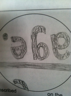
Specifically, in class we were focusing on the "e." The clipping was magnetized 40x and the microscope field of view was 4750 micrometers . The image appeared very grainy, almost as if you were looking at it through a broken piece of glass. The appeared upside down because the microscope has 2 lenses that invert the image and normally your brain would see it right side up but you brain gets confused because the microscope flips it twice.
Blonde Hair & Brown Hair
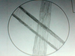
In class, we examined 2 different hairs on the same slide, a blonde hair and a brown hair. We examined the hairs under high power (400x.) Normally, it is extremely difficult to see which hair is on top of the other but in my situation I could tell because there was a distinct border on the edges of the blonde hair (as you can see to the left.)
- When viewing the objects under high power objective, it was very difficult to make the whole image focus. This occurred because when the image is focused it is only focused in the center because the microscope is incapable of focusing on two parts of an image at one time.
- As the magnification increased the field of view increased. This occurred because when the magnification went up the lens got closer to the image and got smaller.
The Skin of an Onion
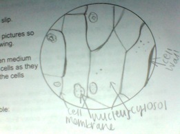
In these observations, we used Lugol's Iodine to color the Onion so that the cells were easier to see. The drawing to the left was made under high power objective (400x.) We also labeled the cell membrane, nucleus, cytosol, and cell wall.
Cheek Cells
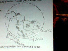
Lastly, I used a clean toothpick to remove some cheek cells from my mouth. I placed the cells on a slide and used water and methylene blue to better see the cells. I observed the cells under high power (400x) and labeled the plasma membrane, nucleus, and cytosol.
- I found it very difficult to tell the difference of the cell wall, cell membrane, and the plasma membrane
- I also felt that the cell stains, such as methylene blue and Lugol's Iodine, made it easier to observe the cells because cells are naturally clear and it would be very difficult to observe a clear specimen on a clear piece of glass.
